What does fact checked mean?
At Healthfully, we strive to deliver objective content that is accurate and up-to-date. Our team periodically reviews articles in order to ensure content quality. The sources cited below consist of evidence from peer-reviewed journals, prominent medical organizations, academic associations, and government data.
- Annals of Surgery: Melanin Spots: Diagnostic Significance in Polyposis
- Annals of Surgery: Melanin Spots: Diagnostic Significance in Polyposis
- PubMed.gov: Late Diagnosis of a Primary Oral Malignant Melanoma: A Case Report.
- PubMed.gov: Late Diagnosis of a Primary Oral Malignant Melanoma: A Case Report.
The information contained on this site is for informational purposes only, and should not be used as a substitute for the advice of a professional health care provider. Please check with the appropriate physician regarding health questions and concerns. Although we strive to deliver accurate and up-to-date information, no guarantee to that effect is made.
What Do Black Spots on Gums Mean?
Most spots found on gums are not serious and result from natural skin pigments, tiny bruises, hardened plaque, pieces of silver fillings under the gum or a vein; but in rare cases they can mean oral malignant melanoma, a form of oral cancer. This disease is often fatal. If you notice any change to your gums, visit a dentist to get a thorough diagnosis.
Calculus
Calculus, also known as tartar, occurs when plaque hardens and, according to Dr. Phyllis Beemsterboer of UCLA, it cannot be removed by simply brushing or flossing. Calculus can range in color from dark yellow to black, and can appear along your gum line in the form of spots; it can lead to bad breath and receding gums. A dentist can remove excess calculus, but regular flossing and brushing will prevent it from occurring. According to the Howard Hughes Medical Institute investigator David M. Kingsley, special toothpastes that combat calculus, containing pyrophosphates, are available 1.
- Calculus, also known as tartar, occurs when plaque hardens and, according to Dr. Phyllis Beemsterboer of UCLA, it cannot be removed by simply brushing or flossing.
Melanin Spots
What are the causes of pale gray and swollen gums?
Learn More
People with dark skin or with dark-skinned ancestry are prone to melanin spots forming on the gums. Melanin is a naturally occurring pigment and is generally harmless, although there are laser treatments that can remove these spots. According to the University of Tennessee College of Medicine, in rare cases melanin spots can be linked to the hereditary disease polyposis 2.
Amalgam Tattoos
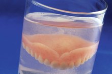
These gum discolorations are quite common and benign. They are formed when particles from silver fillings root themselves in the mouth’s soft tissue. The spots can also occur if you have false teeth, as particles from the metal of a cap are lodged within the gum. They can be black, dark blue or gray and look like a tattoo. According to the Oral Cancer Foundation, they are harmless and can occur over time, or during a dental operation. Although benign, they can be removed by a dentist if you think they look unsightly. Otherwise they are permanent.
- These gum discolorations are quite common and benign.
- The spots can also occur if you have false teeth, as particles from the metal of a cap are lodged within the gum.
Oral Malignant Melanoma
What Are the Causes of a Growth on the Roof of the Mouth?
Learn More
Malignant melanoma is the deadliest form of skin cancer but it is rarely found in the mouth. According to the U.S. National Library of Medicine, oral malignant melanoma accounts for between 0.2 percent and 8 percent of all melanomas. If you smoke, chew tobacco or drink heavily your chances of developing oral melanoma are increased. It starts as a small black spot, generally less than 1mm in diameter, which is actually a tumor that grows, with uneven edges. Malignant melanomas are often fatal, because they are often not discovered until they have reached an advanced stage, by which time survival rate is, on average, two years.
- Malignant melanoma is the deadliest form of skin cancer but it is rarely found in the mouth.
- Malignant melanomas are often fatal, because they are often not discovered until they have reached an advanced stage, by which time survival rate is, on average, two years.
Prevention/Solution
Brushing and flossing regularly will improve your oral hygiene. Avoid chewing tobacco, smoking and drinking excessively. Go for regular dental checks. That way your dentist can detect early signs of disease. If she notices something like oral malignant melanoma before it grows, it could save your life. If you notice any black spots on your gums, get them checked immediately by a dentist.
- Brushing and flossing regularly will improve your oral hygiene.
- That way your dentist can detect early signs of disease.
Related Articles

What are the causes of pale gray and swollen gums?
Learn More

What Are the Causes of a Growth on the Roof of the Mouth?
Learn More

What Are the Causes of Tongue Skin Tags?
Learn More

What Kinds of Growths Form on Aging Skin?
Learn More
Types of Dental Partial Plates
Learn More

Diseases and Disorders of the Tongue
Learn More

Age Spots and Moles
Learn More

How to Remove Tooth Tartar at Home
Learn More

What Causes the Mouth to Look Sunken As We Age?
Learn More

How to Remove Gum Line Stains
Learn More
References
Writer Bio
Based in London, Martin Green has written news, health and sport articles since 2008. His articles have appeared in “Essex Chronicle," “The Journal” and various regional British newspapers. Green holds a Master of Arts in creative writing from Newcastle University and a Bachelor of Arts in English literature.