How to Read Bone Scans
A bone scan is a radiologic technique in which a small amount of a radioactive chemical, also known as a tracer, is injected into the veins 2. This tracer is used to help visualize how fast bones are being broken down and reformed. The radioactive dye will accumulate in areas that have more bone turnover, causing bright spots to appear on the scan 2. Bone scans can be used to identify areas in the bones that have abnormal metabolism, although they are not good for determining the cause of these abnormalities.
If you are experiencing serious medical symptoms, seek emergency treatment immediately.
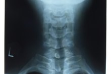
Identify the bones being scanned. Often the bone scan will be labeled, at least in terms of which side is the patient's left and right side, since the scan may be taken from behind or the front of the patient 2. Otherwise, the bones on the scan can be roughly identified by looking at the outline of the organs around the bones.
What Do White Spots on a Bone Scan Indicate?
Learn More
Determine the time frame. Some bone scans consist of just one image. Other bone scans take three different scans over time to look at the bone at different times. Look at the label on the scan to figure out when the scan was taken compared with other scans.
Look for "hot" and "cold" spots. Bone scans use a mildly radioactive dye to measure bone metabolism. "Hot" areas--areas that are abnormally bright on the scan--indicate that there is increased bone turnover in that area, whereas cold areas on the scan--abnormally dark compared with the surrounding bone--indicate that there is less bone breakdown and regrowth in an area 2. Hot spots may be indicative of inflammation or a tumor, whereas cold spots can be a sign of decreased blood flow to an area.
Bone scans are good for detecting abnormalities in bone breakdown and metabolism, but they are not good for determining the cause of these abnormalities. Consequently, further tests, including MRIs, may be necessary to further examine any aberrant bone sections.
Related Articles
References
- Mayo Clinic: Bone Scan
- Medline: Bone Scan
- NIAMS: Osteonecrosis
- Van den wyngaert T, Strobel K, Kampen WU, et al. The EANM practice guidelines for bone scintigraphy. Eur J Nucl Med Mol Imaging. 2016;43(9):1723-38. doi:10.1007/s00259-016-3415-4.
- Hahn S, Heusner T, Kümmel S, et al. Comparison of FDG-PET/CT and bone scintigraphy for detection of bone metastases in breast cancer. Acta Radiol. 2011;52(9):1009-14. doi:10.1258/ar.2011.100507.
- Ohta M, Tokuda Y, Suzuki Y, et al. Whole body PET for the evaluation of bony metastases in patients with breast cancer: comparison with 99Tcm-MDP bone scintigraphy. Nucl Med Commun. 2001;22(8):875-9. doi:10.1097/00006231-200108000-00005.
- Cunha JP. Myoview side effects. Updated August 29, 2019.
- National Library of Medicine. Technetium Tc 99m Bicisate. 2006.
- RadiologiyInfo.org. Bone scan (skeletal scintigraphy). Updated March 1, 2018.
- Brenner, A.; Koshy, J.; Morey, J. et al. The Bone Scan. Sem Nuclear Med. 2012;42(1):11-26. DOI: 10.1053/j.semnuclmed.2011.07.005.
- Tateishi, U.; Gamez, C.; Dawood, S. et al. Bone metastases in patients with metastatic breast cancer: morphologic and metabolic monitoring of response to systemic therapy with integrated PET/CT. Radiology. 2008;247:189-96. DOI: 10.1148/radiol.2471070567.
- U.S. Food and Drug Administration. Highlights of Prescribing Information: Technetium Tc 99m Sulfur Colloid Injection. Silver Spring, Maryland; updated July 2011.
- Wyngaeart, T.; Strobel, K.; Kampen, W. et al. The EANM practice guidelines for bone scintigraphy. Eur J Nucl Med Mol Imaging. 2016;43:1723-38. DOI: 10.1007/s00259-016-3415-4.
Writer Bio
Adam Cloe has been published in various scientific journals, including the "Journal of Biochemistry." He is currently a pathology resident at the University of Chicago. Cloe holds a Bachelor of Arts in biochemistry from Boston University, a M.D. from the University of Chicago and a Ph.D. in pathology from the University of Chicago.
