Kidner Procedure Rehab Protocols
A Kidner procedure is surgery that is performed for an accessory navicular bone 12. The navicular bone is located in the arch of the foot and is attached to the posterior tibial tendon. This architecture helps hold up the arch. An accessory navicular bone is an extra bone located in the instep that causes a flat foot 12. In this condition, the navicular and accessory navicular bones fuse together, and this condition can cause pain 12. Surgical correction is performed only after nonsurgical treatment has failed, and rehabilitation protocols are prescribed by the surgeon.
If you are experiencing serious medical symptoms, seek emergency treatment immediately.
Symptoms and Diagnosis
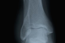
The symptom most associated with accessory navicular is pain in the instep, which can be aggravated by walking 2. If the accessory navicular is to become painful, it will typically happen during the teen years 12. If the accessory navicular does not cause pain, no nonsurgical or surgical treatment is needed 2. Physicians identify the problem when the patient reports pain and typically can confirm the condition with X-rays only.
Kidner Surgical Procedure
Rehabilitation Exercises for Vastus Lateralis
Learn More
The Kidner procedure is a straightforward surgery. To correct the accessory navicular, the surgeon makes a small incision over the extra bone 12. The bone is then detached from the posterior tibial tendon and removed. The tendon is attached to the navicular bone, and the skin incision is closed.
- The Kidner procedure is a straightforward surgery.
- The tendon is attached to the navicular bone, and the skin incision is closed.
General Rehabilitation Protocol
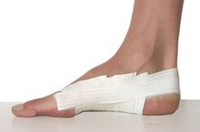
The patient receives a below-knee fiberglass cast, which is worn for approximately three weeks, after which the medical team applies a partial weightbearing walking cast. The patient uses crutches for several days, and a physical therapist can instruct the patient in the proper use of crutches. If sutures are non-absorbable, they are removed in 10 to 14 days following surgery. At approximately four weeks following surgery, the walking cast is removed and the patient starts physical rehabilitation, which consists of a series of stretching exercises to ease tension on the tendon. Normal activities resume in approximately three months.
- The patient receives a below-knee fiberglass cast, which is worn for approximately three weeks, after which the medical team applies a partial weightbearing walking cast.
- At approximately four weeks following surgery, the walking cast is removed and the patient starts physical rehabilitation, which consists of a series of stretching exercises to ease tension on the tendon.
Physical Rehabilitation Protocol
How to Tape a Heel Spur
Learn More
The surgeon determines if physical rehabilitation is necessary following surgery. Physical therapy begins with ice, massage and whirlpool to control pain and swelling and to strengthen the tendon. Therapists often recommend aqua therapy, as water provides a safe environment for the patient to exercise without undue tension on the tendon. As strength improves, the patient takes part in advancing stages of exercise, consisting of stretching the tendon to provide more mobility. Physical therapy helps improve range of motion and ensures that the patient is walking normally. Once normal movement and strength resume, physical therapy ends and the patient continues the exercise program at home to ensure that the tendon remains flexible.
- The surgeon determines if physical rehabilitation is necessary following surgery.
- As strength improves, the patient takes part in advancing stages of exercise, consisting of stretching the tendon to provide more mobility.
Related Articles

Rehabilitation Exercises for Vastus Lateralis
Learn More
How to Tape a Heel Spur
Learn More
Why Does My Achilles Tendon Hurt?
Learn More

Knee Cap Pain & Swelling
Learn More
How Do I Treat a Bone Chip?
Learn More
Unexplained Shooting Pain in My Shins
Learn More
Fractured Ankle & Numbness
Learn More

Iliopsoas Tendon Pain When Walking
Learn More
Taping Techniques for Morton's Neuroma
Learn More
How to Rehab a Suspensory Strain
Learn More
References
- Podiatry Today: How to Conquer the Accessory Navicular Bone
- EOrthopod: Accessory Navicular Problems
- Santa Rosa Orthopedics: Accessory Navicular Problems
- Gross CE, Nunley JA. Navicular Stress Fractures. Foot Ankle Int. 2015;36(9):1117-22. doi:10.1177/1071100715600495
- Fowler JR, Gaughan JP, Boden BP, Pavlov H, Torg JS. The non-surgical and surgical treatment of tarsal navicular stress fractures. Sports Med. 2011;41(8):613-9. doi:10.2165/11590670-000000000-00000
- Miller TL, Jamieson M, Everson S, Siegel C. Expected Time to Return to Athletic Participation After Stress Fracture in Division I Collegiate Athletes. Sports Health. 2018;10(4):340-344 doi:10.1177/1941738117747868
- Shindle MK, et al. "Stress Fractures About the Tibia, Foot, and Ankle" J Am Acad Orthop Surg. 2012 Mar;20(3):167-76.
Writer Bio
Lynda Lanford began her writing career in the technological arena in 1989, working for such organizations as National Computer Systems and KnowledgeNet. She has also worked in medical transcription. This combination of experience has led to a strong interest and capacity for writing medical topics in everyday language. Lanford earned an Associate of Arts degree from Glendale College with a journalism emphasis.