What does fact checked mean?
At Healthfully, we strive to deliver objective content that is accurate and up-to-date. Our team periodically reviews articles in order to ensure content quality. The sources cited below consist of evidence from peer-reviewed journals, prominent medical organizations, academic associations, and government data.
The information contained on this site is for informational purposes only, and should not be used as a substitute for the advice of a professional health care provider. Please check with the appropriate physician regarding health questions and concerns. Although we strive to deliver accurate and up-to-date information, no guarantee to that effect is made.
Vein Ablation Complications
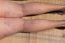
Varicose veins can be extremely disfiguring and distressing for both women and men, though they are much more common in women 3. This condition occurs when the valves and walls of veins in the legs, ankles and feet weaken leading to engorged and twisted veins that bulge from the surface of the skin. The Mayo Clinic advises that varicose veins can be asymptomatic and only a cosmetic concern for some individuals while others experience painful aching, burning, itching and bleeding symptoms 23. There are several treatments for varicose veins including laser ablation procedures such as endovenous laser treatment or ELST 23. Other treatments include sclerotherapy in which a solution is injected to scar and close the vein. Varicose vein ablation treatments are effective, but can cause complications 2.
Bruising and Bleeding
Procedures for the ablation of varicose veins include endovenous laser treatment or EVLT and other laser vein treatments 123. While some laser treatment target smaller varicose veins from above the skin, The Vein Institute of Toronto explains that EVLT is slightly more invasive 13. This ablation procedure requires a small incision in the leg to insert a catheter that is threaded into the varicosed vein. The catheter then gives off laser energy that scars and seals the damaged vein. The closed vein is removed passively by the body in several weeks. These procedures can lead to bleeding and bruising at the site of the incision and along the vein that has been treated. Bleeding from the incision does not last longer than 30 minutes or more after the treatment but bruising can last for as long as a few weeks.
- Procedures for the ablation of varicose veins include endovenous laser treatment or EVLT and other laser vein treatments 1.
- Bleeding from the incision does not last longer than 30 minutes or more after the treatment but bruising can last for as long as a few weeks.
Pain
Complications of Endovenous Laser Treatment
Learn More
Laser ablation procedures are painful and are usually performed with local anesthesia. However, once the numbing wears off shortly after the treatment, pain, aching and burning can occur in the treated leg for a few days or longer. In some cases a temporary loss of sensation due to damaged nerves may also occur along the area of the treated vein.
Scarring
Laser ablation procedures for veins can also cause scarring. The Mayo Clinic warns that there is a small risk of scarring at the point of incision where the cather is inserted into the leg. In surface laser vein procedures, there is a risk of minor to moderate burns and scars where the laser heat penetrates the skin to reach the vein. In most cases, if these procedures are performed by professionals the risk of scarring is very minimal.
- Laser ablation procedures for veins can also cause scarring.
- In surface laser vein procedures, there is a risk of minor to moderate burns and scars where the laser heat penetrates the skin to reach the vein.
Recurrence
What to Expect From a 40% Glycolic Peel
Learn More
Vein ablation procedures with lasers may require additional treatments if the varicose is not completely removed in one treatment 12. According to The Vein Institute of Toronto, endovenous laser therapy has a recurrence rate of only 5 percent while surface laser treatments that must use lower energy lasers to avoid damaging the skin have a higher recurrence rate and may need additional treatments 1.
Related Articles

Complications of Endovenous Laser Treatment
Learn More
What to Expect From a 40% Glycolic Peel
Learn More

How to Remove Age Spots & Brown Spots by Freezing
Learn More

Bad Side Effects of Juvederm
Learn More
Popliteal Aneurysm Symptoms
Learn More
Betaderm Side Effects
Learn More

Blood Clot After Surgery Symptoms
Learn More
Derma Filler Side Effects
Learn More

How to Get Rid of Stitch Scars on the Face
Learn More
Care of Skin After Cryosurgery
Learn More
References
- The Vein Institute of Toronto: Varicose Vein Treatment
- Mayo Clinic: Varicose Veins Treatments
- Mayo Clinic: Varicose Veins
- Raetz J, Wilson M, Collins K. Varicose veins: Diagnosis and treatment. Am Fam Physician. 2019;99(11):682-688.
- Critello CD, Pullano SA, Matula TJ, De Franciscis S, Serra R, Fiorillo AS. Recent developments on foaming mechanical and electronic techniques for the management of varicose veins. Expert Rev Med Devices. 2019;16(11):931-940. doi:10.1080/17434440.2019.1682549
- Epstein D, Onida S, Bootun R, Ortega-Ortega M, Davies AH. Cost-effectiveness of current and emerging treatments of varicose veins. Value Health. 2018;21(8):911-920. doi:10.1016/j.jval.2018.01.012
- American Academy of Dermatology. Leg veins: Why they appear and how dermatologists treat them.
Writer Bio
Noreen Kassem is a hospital doctor and a medical writer. Her articles have been featured in "Women's Health," "Nutrition News," "Check Up" and "Alive Magazine." Kassem also covers travel, books, fitness, nutrition, cooking and green living.