What does fact checked mean?
At Healthfully, we strive to deliver objective content that is accurate and up-to-date. Our team periodically reviews articles in order to ensure content quality. The sources cited below consist of evidence from peer-reviewed journals, prominent medical organizations, academic associations, and government data.
- Mayo Clinic: Age Spots Treatments and Drugs
- Cleveland Clinic: Incontinentia Pigmenti
- Cleveland Clinic: Sun Exposure and Skin Cancer
The information contained on this site is for informational purposes only, and should not be used as a substitute for the advice of a professional health care provider. Please check with the appropriate physician regarding health questions and concerns. Although we strive to deliver accurate and up-to-date information, no guarantee to that effect is made.
Skin Discoloration on the Forehead
Discolorations on the forehead are not uncommon. In fact, it is likely you will develop one at some point in your life, either as a temporary discoloration caused by sun exposure, or through a more permanent darkening or lightening of the skin. Discolorations can be uncomfortable and embarrassing to some people, but in most cases there are ways to treat them.
Types
One type of discoloration that is temporary in nature is a darkening due to sun exposure. The forehead can receive direct sunlight, particularly if it is uncovered and unprotected, and if the exposure is too long it can lead to burning of the skin. This can cause reddening of the skin and later darkening as a result of melanin production. You can also develop white scaling and flaking of the skin as it heals and sheds damaged layers. Darkened spots can also occur either as freckles or larger spots of coagulated melanin, such as birthmarks, flat moles and age spots.
- One type of discoloration that is temporary in nature is a darkening due to sun exposure.
- The forehead can receive direct sunlight, particularly if it is uncovered and unprotected, and if the exposure is too long it can lead to burning of the skin.
Dark Spot Treatments
Dark Pigmentation in the Elbows, Ankles and Inner Thigh
Learn More
Chemical peels are available to remove the top layers of your skin and try to replaced damaged pigment-producing cells with cells that function normally. It can also help to remove accumulations that occur naturally, as in the case of birth marks and liver spots. Other skin bleaching treatments can sometimes be effective. Dermabrasion, cryotherapy and laser therapy are all treatments that can help remove some skin discolorations, according to MayoClinic.com.
- Chemical peels are available to remove the top layers of your skin and try to replaced damaged pigment-producing cells with cells that function normally.
- Dermabrasion, cryotherapy and laser therapy are all treatments that can help remove some skin discolorations, according to MayoClinic.com.
Sunburn Treatments
Sunburns heal on their own, but you can minimize damage to your skin -- as well as the pain experienced -- with some simple treatments. First and foremost, shield your forehead from the sun by wearing a hat and/or applying sunscreen. Also moisturize the skin at least twice a day, which can also be done with a moisturizing lotion or by applying a moisturizing sunscreen. Apply aloe vera gel can also soothe the sunburn, making it less painful and less likely to peel.
- Sunburns heal on their own, but you can minimize damage to your skin -- as well as the pain experienced -- with some simple treatments.
- Also moisturize the skin at least twice a day, which can also be done with a moisturizing lotion or by applying a moisturizing sunscreen.
Considerations
Tyrosine & Vitiligo
Learn More
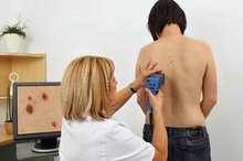
Most discolorations are harmless, but there are some cases where you should visit a doctor. If a skin discoloration like a flat mole appears to change tone, have it examined by a doctor to determine whether it may be a warning sign of cancer or other problems. If your skin blisters from overexposure to sunlight, you might also want to have your skin treated and cleansed.
Prevention/Solution
Some types of spots cannot be avoided, such as the development of liver spots. But many types of skin discolorations can be caused by sun exposure. Limiting your forehead's exposure to the sun by wearing hats, scarves or other head coverings, and using sunscreen to protect it when your head is exposed, can reduce your risk of skin damage both in the short term as well as over time 3.
Related Articles

Dark Pigmentation in the Elbows, Ankles and Inner Thigh
Learn More
Tyrosine & Vitiligo
Learn More

Types of Skin Cancer on the Nose
Learn More
Age Spots and Moles
Learn More
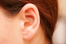
Earlobe Wart
Learn More
Actinic Keratosis Vs. Seborrheic Keratosis
Learn More

Why Is a Mole Raising Up on Skin?
Learn More

Early Signs of Skin Cancer and Penile Cancer
Learn More
Vitamin E for Skin Discoloration
Learn More

Hyperpigmentation While on Birth Control
Learn More
References
- Mayo Clinic: Age Spots Treatments and Drugs
- Cleveland Clinic: Incontinentia Pigmenti
- Cleveland Clinic: Sun Exposure and Skin Cancer
- Vashi NA, de Castro Maymone MB, Kundu RV. Aging differences in ethnic skin. J Clin Aesthet Dermatol. 2016;9(1):31-38.
- American Osteopathic College of Dermatology. Hyperpigmentation.
- Bandyopadhyay D. Topical treatment of melasma. Indian J Dermatol. 2009;54(4):303-309. doi:10.4103/0019-5154.57602
- Kang SJ, Choi BR, Lee EK, et al. Inhibitory effect of dried pomegranate concentration powder on melanogenesis in B16F10 melanoma cells; involvement of p38 and PKA signaling pathways. Int J Mol Sci. 2015;16(10):24219-42. doi:10.3390/ijms161024219
- Parveen R, Akhtar N, Mahmood T. Topical microemulsion containing Punica granatum extract: its control over skin erythema and melanin in healthy Asian subjects. Postepy Dermatol Alergol. 2014;31(6):351-5. doi:10.5114/pdia.2014.47117
- Kim E, Hwang K, Lee J, et al. Skin protective effect of epigallocatechin gallate. Int J Mol Sci. 2018;19(1). doi:10.3390/ijms19010173
- Kim YC, Choi SY, Park EY. Anti-melanogenic effects of black, green, and white tea extracts on immortalized melanocytes. J Vet Sci. 2015;16(2):135-43. doi:10.4142/jvs.2015.16.2.135
- Zhou BR, Ma LW, Liu J, et al. Corrigendum to "protective effects of soy oligopeptides in ultraviolet B-induced acute photodamage of human skin". Oxid Med Cell Longev. 2018;2018:3871280. doi:10.1155/2016/5846865
- Adhikari D, Panthi VK, Pangeni R, Kim HJ, Park JW. Preparation, characterization, and biological activities of topical anti-aging ingredients in a Citrus junos callus extract. Molecules. 2017;22(12). doi:10.3390/molecules22122198
Writer Bio
Jonathan Croswell has spent more than five years writing and editing for a number of newspapers and online publications, including the "Omaha World-Herald" and "New York Newsday." Croswell received a Bachelor of Arts degree in English from the University of Nebraska and is currently pursuing a Master's of Health and Exercise Science at Portland State University.