What does fact checked mean?
At Healthfully, we strive to deliver objective content that is accurate and up-to-date. Our team periodically reviews articles in order to ensure content quality. The sources cited below consist of evidence from peer-reviewed journals, prominent medical organizations, academic associations, and government data.
The information contained on this site is for informational purposes only, and should not be used as a substitute for the advice of a professional health care provider. Please check with the appropriate physician regarding health questions and concerns. Although we strive to deliver accurate and up-to-date information, no guarantee to that effect is made.
How Long Does It Take Blood to Circulate?
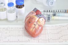
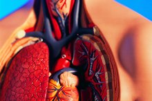
The Heart
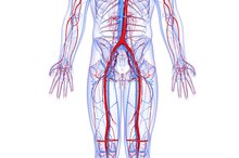
Blood moves through the arteries and veins in only one direction. The opening and closing of valves in the heart’s chambers work in such a way to make certain that blood is carried throughout the body in a circle known as circulation. The heart pumps blood through vessels transporting oxygen and nutrients to all the parts of the body. Larger blood vessels, called arteries, carry oxygen rich blood from the heart. Blood then flows from the smaller arteries in the body into even smaller blood vessels called capillaries, where oxygen is dropped off and carbon dioxide is picked up. For the return trip, blood in the capillaries flows into small veins, which flow into larger veins that lead back to the heart. Blood travels from there to the lungs, where it releases carbon dioxide, a cellular waste product.
If you are experiencing serious medical symptoms, seek emergency treatment immediately.
- Blood moves through the arteries and veins in only one direction.
- For the return trip, blood in the capillaries flows into small veins, which flow into larger veins that lead back to the heart.
Heart Rate
Blood takes less time to circulate when you are active or exercising, as your heart rate decreases when you are resting 1. The healthy adult heart beats approximately 65 to 75 beats per minute. With each beat, the heart pumps an average of 60 to 70 milliliters of blood, or about five liters per minute. The American Heart Association points out that based on an average of 100,000 beats each day, the heart pumps out 2,000 gallons of blood. According to information published by the Franklin Institute, it is estimated that if you were to line up all the blood vessels in the human body from end to end, they would circle the Earth four times. That means that when you add them up the body of an adult contains 100,000 miles of arteries, veins and capillaries. The blood vessels in a child’s body would be more than 60,000 miles long.
- Blood takes less time to circulate when you are active or exercising, as your heart rate decreases when you are resting 1.
- The blood vessels in a child’s body would be more than 60,000 miles long.
Circulation
What Are the Largest Blood Vessels in the Body?
Learn More
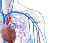
Since the body requires a steady supply of blood to keep the organs functioning properly, it is essential for the heart to keep pumping blood to each of the body’s cells 2. The length of time it takes blood to circulate throughout the body depends on a person’s size, overall physical condition and health, age and heart rate 12. The right side of the heart pushes the blood returning through the veins into the large pulmonary artery that carries blood to the lungs where it releases carbon dioxide and receives more oxygen 2. Blood is then pumped into the pulmonary veins, which return it to the left side of the heart where it begins the circulation process again 2. It takes the heart less than one minute to pump blood to every cell in the body 2. In fact, six quarts of blood can fully circulate throughout the body at least three times within that minute 1. That totals thousands of round trips each day.
Related Articles
What Are the Largest Blood Vessels in the Body?
Learn More
The 4 Parts of the Cardiovascular System
Learn More
How Do the Walls of the Atria & Ventricles Differ?
Learn More
What Are the Three Major Parts of the Cardiovascular System?
Learn More
How Do the Walls of the Atria & Ventricles Differ?
Learn More

What Causes a Pulse in an Artery?
Learn More

How the Heart & Lungs Work During Exercise
Learn More
Differences Between Blood Vessels & Lymph Vessels
Learn More
Parts of the Lungs
Learn More
What Are the Functions of Simple Squamous Epithelial Cells?
Learn More
References
- How Does Blood Circulate
- Heart, How It Works
- National Institute on Aging: Aging Hearts and Arteries.
Writer Bio
Amber Keefer has more than 25 years of experience working in the fields of human services and health care administration. Writing professionally since 1997, she has written articles covering business and finance, health, fitness, parenting and senior living issues for both print and online publications. Keefer holds a B.A. from Bloomsburg University of Pennsylvania and an M.B.A. in health care management from Baker College.