What does fact checked mean?
At Healthfully, we strive to deliver objective content that is accurate and up-to-date. Our team periodically reviews articles in order to ensure content quality. The sources cited below consist of evidence from peer-reviewed journals, prominent medical organizations, academic associations, and government data.
The information contained on this site is for informational purposes only, and should not be used as a substitute for the advice of a professional health care provider. Please check with the appropriate physician regarding health questions and concerns. Although we strive to deliver accurate and up-to-date information, no guarantee to that effect is made.
How to Read the Lines on a Heart Monitor
You may not be a cardiologist, but you can glean a certain amount of information from the lines on an electrocardiogram (EKG) 1. Still, it's important to leave the true reading and diagnosis to trained healthcare providers whose profession it is to detect abnormal sinus rhythms.
Deciphering peaks and valleys
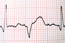
Compare your EKG to the normal sample (see ecglibrary.com/norm.html). The vertical lines on the paper cut the waves into five sections denoted with letters PQRST. P is the small hump before the dip, Q is the tiny dip before the spike, R is the spike, S is the dip after the spike and T is the low spike after the dip.
How to Read an EKG for Dummies
Learn More
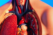
The beginning P-wave width represents the atria (upper part of the heart that receives the blood) with a time interval labelled as PR. Its time is 0.12 to 0.20 seconds. The next section, QRS, is associated with the ventricle (the lower part of the heart) and has a time interval of 0.04 to 0.10 seconds. The QT has an time interval of less than 0.02 seconds. Circle these intervals.
Compare the height of the P and T humps and R-spike and their sharpness. Also compare the width of them to the normal print out. Slow sinus rhythms called bradycardia have fewer, wider peaks and valleys. Faster sinus rhythms called tachycardia have more and thinner peaks and valleys.
How to Count the Boxes on the EKG Paper to Determine Rate
Learn More
Compare the dips-Q and S to the normal sinus rhythm print out. Deep or elevated ST section are associated with heart attack. Depressed or flatter waves are associated with blockages in the heart.
Do not be overly concerned if your EKG shows flat lines in between peaks and valleys--this is normal. However, extended flat lines most likely indicate that the electrodes are not picking up any transmission from the heart. Call your doctor immediately
Tips
Medications can influence the lines (Digoxin, for example). Eating or drinking before the test can alter heart rate. Exercise can alter heart rate. Placement of the electrodes can affect the reading.
Warnings
Just because you have noticed abnormalities on the lines doesn't mean that you have heart disease. Readings that seem to indicate problems when there's no history of heart disease, obesity or chest pain are most likely a fluke. Always consult your physician before and after any test.
- Compare your EKG to the normal sample (see ecglibrary.com/norm.html).
- The next section, QRS, is associated with the ventricle (the lower part of the heart) and has a time interval of 0.04 to 0.10 seconds.
Related Articles
How to Read an EKG for Dummies
Learn More
How to Count the Boxes on the EKG Paper to Determine Rate
Learn More

How to Read an Abnormal EKG
Learn More
What Are the Four Parts of the EKG Machine?
Learn More
How to Hear Your Own Heartbeat Without a Stethoscope
Learn More
Normal Pulse Rate at 13 Years Old
Learn More
Can You Feel Your Heartbeat by Touching Your Chest?
Learn More
How to Feel a Pulse Between the Ribs
Learn More
How to Read a Fetal Monitor Strip
Learn More

8 High-Tech Gadgets to Keep Your Heart Healthy
Learn More
References
Tips
- Medications can influence the lines (Digoxin, for example).
- Eating or drinking before the test can alter heart rate.
- Exercise can alter heart rate.
- Placement of the electrodes can affect the reading.
Warnings
- Just because you have noticed abnormalities on the lines doesn't mean that you have heart disease. Readings that seem to indicate problems when there's no history of heart disease, obesity or chest pain are most likely a fluke. Always consult your physician before and after any test.
Writer Bio
Bernadette Sukley's work has been published in "Natural Health," "Sports Illustrated for Women," "Men's Health" and "Swimmer" magazines as well as local magazines and newspapers. She's been fact checking, writing and editing for over 20 years. Sukley specializes in health and lifestyle topics.