What does fact checked mean?
At Healthfully, we strive to deliver objective content that is accurate and up-to-date. Our team periodically reviews articles in order to ensure content quality. The sources cited below consist of evidence from peer-reviewed journals, prominent medical organizations, academic associations, and government data.
The information contained on this site is for informational purposes only, and should not be used as a substitute for the advice of a professional health care provider. Please check with the appropriate physician regarding health questions and concerns. Although we strive to deliver accurate and up-to-date information, no guarantee to that effect is made.
What Are the Causes of Incomplete Mammogram Results?
Don't panic if you've been told your mammogram results are incomplete. This usually means the radiologist needs more information to make the most accurate assessment of your results. The radiologist may request further studies or copies of previous studies for comparison purposes. After examining all information available, your mammogram will be categorized according to BI-RADS, or the Breast Imaging Reporting and Database System used by the American College of Radiology.
Function
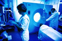
Mammograms are low dose X-rays of breast tissue used to detect breast cancer 1. The National Cancer Institute recommends mammograms be obtained every one to two years for women age 40 and over 1. You may be asked to obtain a baseline mammogram sooner than age 40 so that it may be compared with later mammograms. Women with a history of breast cancer or a family history of breast cancer may need mammograms sooner than age 40 and more often than every one to two years 1.
Types of Mammograms
What Is the Difference Between a Regular Mammogram & a Bilateral Mammogram?
Learn More
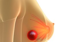
A screening mammogram is done for women with no signs or symptoms of breast cancer. These x-rays can detect breast lesions which cannot yet be seen or felt by physical exam. Diagnostic mammograms are done when signs or symptoms of breast pathology are present, including lumps, nipple discharge, pain or tenderness, thickened skin over the breast, or a change in the breasts size or shape. A diagnostic mammogram may also be done as a follow-up if any abnormalities were found on your screening mammogram, or if the radiologist needs more information.
- A screening mammogram is done for women with no signs or symptoms of breast cancer.
- A diagnostic mammogram may also be done as a follow-up if any abnormalities were found on your screening mammogram, or if the radiologist needs more information.
Digital Mammography
Digital mammograms are different from conventional mammograms in that the x-rays taken are stored directly onto a computer and not on film. According to the National Cancer Institute, because:
- digital images can be manipulated
- magnified
- or enhanced
- subtle differences between normal
- abnormal breast tissue may be more easily detected than with conventional film mammography 1
Digital mammography may be appropriate for pre- or perimenopausal women with dense breasts. However, both types of mammography are equally able to detect breast cancer.
Assessment
Why Is Creatine Level Important for a Brain MRI?
Learn More
BI-RADS provides standardized assessment of breast imaging, whether the imaging is a mammogram, an ultrasound, or a magnetic resonance imaging scan. If your assessment is complete, your imaging will be assigned to one of six categories: \" 1) negative, 2) benign finding, 3) probably benign finding--an initial short-interval follow-up suggested, 4) suspicious abnormality--a biopsy should be considered, 5) highly suggestive of malignancy--appropriate action should be taken, or 6) known biopsy-proven malignancy--appropriate action should be taken.\"
Incomplete Result
If your doctor determines that more information is needed before determining what category to assign your imaging, it will be assigned to BI-RADS category 0 (zero): \"Assessment is incomplete--need additional imaging evaluation and/or prior mammograms for comparison.\" According to the National Women's Health Information Center, if you have dense or fibrous breasts, you may need further evaluation with breast ultrasound or MRI 1. If you've had other breast imaging in the past, your radiologist may want to view the images and reports to compare them to the current X-ray, in order to make the most accurate assessment possible.
Related Articles

What Is the Difference Between a Regular Mammogram & a Bilateral Mammogram?
Learn More
Why Is Creatine Level Important for a Brain MRI?
Learn More

A Breast Lump in a Male
Learn More

How to Determine Low or High FSH Levels
Learn More

What Food or Drink Can Raise Your PSA Levels?
Learn More
What Tests Are Done at the Gynecologist?
Learn More
Characteristics of Breast Cancer Lumps
Learn More

Soy Protein & Male Breasts
Learn More

Differences between Malignant Melanoma and a Normal Mole
Learn More

Early Signs of Skin Cancer and Penile Cancer
Learn More
References
- Mammograms—National Cancer Institute
- Løberg M, Lousdal ML, Bretthauer M, Kalager M. Benefits and harms of mammography screening. Breast Cancer Res. 2015;17(1):63. Published 2015 May 1. doi:10.1186/s13058-015-0525-z
- Centers For Disease Control and Prevention. Breast Cancer Screening Guidelines for Women. Breast Cancer. Published November 5, 2018.
- Koo MM, von Wagner C, Abel GA, McPhail S, Rubin GP, Lyratzopoulos G. Typical and atypical presenting symptoms of breast cancer and their associations with diagnostic intervals: Evidence from a national audit of cancer diagnosis. Cancer Epidemiol. 2017;48:140–146. doi:10.1016/j.canep.2017.04.010
- Yalaza M, İnan A, Bozer M. Male Breast Cancer. J Breast Health. 2016;12(1):1–8. Published 2016 Jan 1. doi:10.5152/tjbh.2015.2711
- Sinclair N, Littenberg B, Geller B, Muss H. Accuracy of Screening Mammography in Older Women. American Journal of Roentgenology. 2011;197(5):1268-1273. doi:10.2214/ajr.10.5442
- Jain M, Jain A, Hyzy MD, Werth G. FAST MRI breast screening revisited. Journal of Medical Imaging and Radiation Oncology. 2016;61(1):24-28. doi:10.1111/1754-9485.12502
- Amano G. MRI Accurately Depicts Underlying DCIS in a Patient with Pagets Disease of the Breast Without Palpable Mass and Mammography Findings. Japanese Journal of Clinical Oncology. 2005;35(3):149-153. doi:10.1093/jjco/hyi044
- Yeh ED, Jacene HA, Bellon JR. What Radiologists Need to Know about Diagnosis and Treatment of Inflammatory Breast Cancer: A Multidisciplinary Approach. RadioGraphics. 2013;33(7):2003-2017. doi:10.1148/rg.337135503
- Horsley RK, Kling JM, Vegunta S, Lorans R, Temkit HH, Patel BK. Baseline Mammography: What Is It and Why Is It Important? A Cross-Sectional Survey of Women Undergoing Screening Mammography. Journal of the American College of Radiology. 2019;16(2):164-169. doi:10.1016/j.jacr.2018.07.002
- Juanpere S, Perez E, Huc O, Motos N, Pont J, Pedraza S. Imaging of breast implants—a pictorial review. Insights into Imaging. 2011;2(6):653-670. doi:10.1007/s13244-011-0122-3
- Akram M, Iqbal M, Daniyal M, Khan AU. Awareness and current knowledge of breast cancer. Biol Res. 2017;50(1):33. Published 2017 Oct 2. doi:10.1186/s40659-017-0140-9
- Vreugdenburg TD, Willis CD, Mundy L, Hiller JE. A systematic review of elastography, electrical impedance scanning, and digital infrared thermography for breast cancer screening and diagnosis. Breast Cancer Research and Treatment. 2013;137(3):665-676. doi:10.1007/s10549-012-2393-x
- Lee CI, Chen LE, Elmore JG. Risk-based Breast Cancer Screening: Implications of Breast Density. Med Clin North Am. 2017;101(4):725–741. doi:10.1016/j.mcna.2017.03.005
- Miglioretti DL, Lange J, van den Broek JJ. Radiation-Induced Breast Cancer Incidence and Mortality From Digital Mammography Screening: A Modeling Study. Ann Intern Med. 2016;164(4):205–214. doi:10.7326/M15-1241
- Rose MG. Oncology in Primary Care. Wolters Kluwer Health: Lippincott Williams & Wilkins; 2013.
- Lee NC, Wong FL, Jamison PM. Implementation of the National Breast and Cervical Cancer Early Detection Program: the beginning. Cancer. 2014;120 Suppl 16(0 16):2540–2548. doi:10.1002/cncr.28820
- de Groot JE, Broeders MJ, Grimbergen CA, den Heeten GJ. Pain-preventing strategies in mammography: an observational study of simultaneously recorded pain and breast mechanics throughout the entire breast compression cycle. BMC Womens Health. 2015;15:26. doi:10.1186/s12905-015-0185-2
- Sharp PC, Michielutte R, Freimanis R, Cunningham L, Spangler J, Burnette V. Reported Pain Following Mammography Screening. Archives of Internal Medicine. 2003;163(7):833. doi:10.1001/archinte.163.7.833
- Karliner LS, Patricia Kaplan C, Juarbe T, Pasick R, Pérez-Stable EJ. Poor patient comprehension of abnormal mammography results. J Gen Intern Med. 2005;20(5):432–437. doi:10.1111/j.1525-1497.2005.40281.x
- Rao AA, Feneis J, Lalonde C, Ojeda-Fournier H. A Pictorial Review of Changes in the BI-RADS Fifth Edition. RadioGraphics. 2016;36(3):623-639. doi:10.1148/rg.2016150178
- Seely JM, Alhassan T. Screening for breast cancer in 2018-what should we be doing today?. Curr Oncol. 2018;25(Suppl 1):S115–S124. doi:10.3747/co.25.3770
- American Cancer Society. Limitations of Mammograms. Atlanta, Georgia; updated 10/09/17.
- Harris, J., Lippman M., Morrow, M., Osborne, C., eds. (2014) Diseases of the Breast. 5th ed. Philadelphia, PA: Lippincott, Williams & Wilkins.
- Miglioretti, D., Lange, J., van den Broek, J. et al. Radiation-Induced Breast Cancer Incidence and Mortality from Digital Mammography Screening. Ann Int Med. 2016. 164(4):205-14. DOI: 10.7326/m15-1241.
- U.S. Food and Drug Administration. Breast Cancer Screening: Thermogram No Substitute for Mammogram. Silver Spring, Maryland; updated 10/27/17.
Writer Bio
Based in Arizona, Kira Jaines writes health/fitness and travel articles, volunteers with Learning Ally and travels throughout the Southwest. She has more than 16 years of experience in transcribing and editing medical reports. Jaines holds a Bachelor of Arts in telecommunications and journalism from Northern Arizona University.